Oblique plane microscopy (OPM)
napari-lattice can be used for processing oblique plane microscopy (OPM) and scanned oblique plane illumination (SOPi) data. The default parameters work for the Zeiss Lattice Lightsheet 7 (LLS7). The deskew is parameter-driven — you set the skew axis, deskew angle, whether to apply the coverslip rotation, and whether to invert the scan direction. Choosing those values to match a different acquisition geometry enables napari-lattice to process data from different systems.
This example deskews two published OPM datasets. The two parameters that make an
acquisition "OPM" are usually to do with coverslip_rotation and invert_scan_direction,
and of course different angles and voxel sizes.
Data and configs
The two datasets below — raw image, deskewed output, and the lls-pipeline config
that produced it — are published with the manuscript on Zenodo as
Supplementary_opm_data.zip. The file paths in the configs are placeholders; point
them at your own copies.
The key setting: coverslip rotation
By default napari-lattice applies a coverslip rotation — after shearing, it rotates the deskewed volume by the deskew angle. This is the correct output geometry for the Zeiss LLS7, where it leaves the specimen level with the coverslip.
Many OPM/SOPi systems need the opposite — the volume should not be rotated, because the shear alone already brings the specimen plane level with the coverslip. For these you turn the coverslip rotation off:
| Setting | Plugin | CLI | What it does |
|---|---|---|---|
| Coverslip rotation on (default) | Coverslip Rotation ticked |
omit the flag (or --coverslip-rotation) |
Standard deskew (cle.deskew_y/cle.deskew_x) then rotate by the deskew angle. Coverslip-level for Zeiss LLS7. |
| Coverslip rotation off | Coverslip Rotation unticked |
--no-coverslip-rotation |
Shear-only deskew, no rotation. Coverslip-level for many OPM/SOPi systems. |
This is a geometry choice, not a quality setting
Neither value is "better" — the right one depends on how your microscope acquires the data. If a deskew looks sheared, tilted, or stretched at the light-sheet angle instead of lying flat, the coverslip-rotation setting is the first thing to flip. Both OPM datasets below use coverslip rotation off.
See Coverslip-frame deskew (--no-coverslip-rotation)
for the reference details.
TIP: In the plugin, Quick Deskew gives a fast preview so you can check the orientation before committing to a full run.
The second setting: scan-direction (Z) inversion
Different microscopes scan the sample in opposite directions. On some oblique-plane systems the stage or galvo scans the opposite way to the Zeiss LLS, so the deskewed volume comes out mirrored along the scan (Z) axis.
invert_scan_direction reverses the order of the planes along the scan Z axis before
deskewing, correcting this handedness. Both datasets below need it:
| Setting | Plugin | CLI | What it does |
|---|---|---|---|
| Invert scan direction | Invert Scan Direction ticked |
--invert-scan-direction |
Reverse plane order along the scan (Z) axis before deskewing. Default is off (Zeiss LLS behaviour). |
If a coverslip-rotation-corrected deskew is still flipped top-to-bottom along the depth
axis, this is the setting to change. See
Flipping the scan direction (--invert-scan-direction).
Datasets
| Folder | Sample | Reference |
|---|---|---|
brain_organoid |
4×-expanded brain organoid, direct-view oblique plane microscopy | Lamb et al., Optica 12, 469–472 (2025); raw data 10.6084/m9.figshare.28324301 |
thy1_eGFP |
Uncleared coronal Thy1-GFP mouse brain section, scanned oblique plane illumination (SOPi) | Kumar & Kozorovitskiy, Opt. Lett. 44, 1706–1709 (2019); raw data zenodo.org/records/5088089 |
Both are Y-skew, 45° acquisitions with coverslip rotation off and scan direction inverted. They differ only in pixel size.
Option A — in the napari plugin
-
Drag the raw
.tifinto napari. It loads as an image layer — here thethy1_eGFPSOPi dataset, shown in green. Open the Lattice Lightsheet Analysis plugin (Plugins → napari-lattice) and, on the 1. Deskew tab, select the layer underImage Layer(s) to Deskew.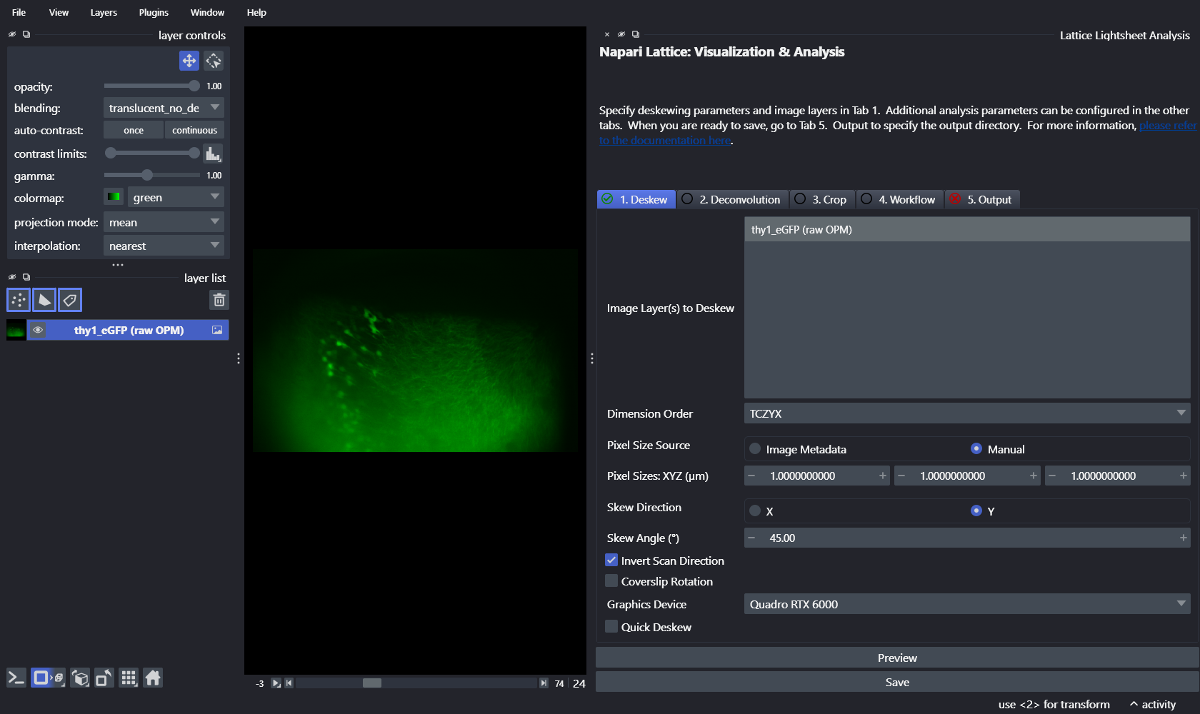
-
Enter the acquisition metadata for the dataset. Set
Pixel Size Sourceto Manual, fill in the pixel sizes and skew angle, choose the skew direction, and — the two settings that make this an OPM run — tickInvert Scan Directionand leaveCoverslip Rotationunticked.Parameter brain_organoidthy1_eGFPdx (µm) 1.04 1 dy (µm) 1.04 1 dz (µm) 2.0 1 Deskew angle 45 45 Skew Direction Y Y Coverslip Rotation off off Invert Scan Direction on on The Deskew tab for the
thy1_eGFPdataset should look like this — note the tickedInvert Scan Directionand the untickedCoverslip Rotation: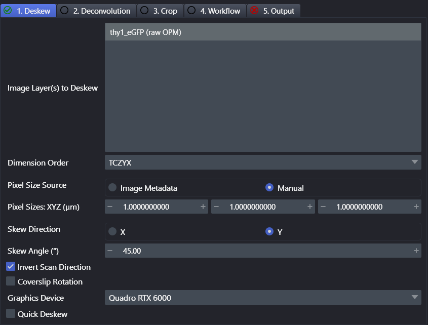
-
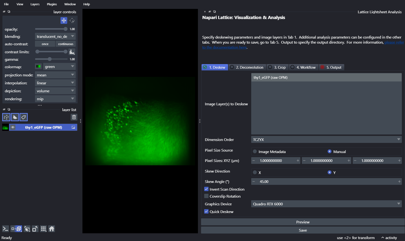
Tick Quick Deskew for a fast preview of the orientation before committing to a full run.
-
When the preview looks right, switch to the 5. Output tab to save. Choose a
Save Format(tiffwrites a compressed OME-TIFF), pick aSave Directory, and adjust theSave Suffix, channel and time ranges if needed. Click Save to run the deskew and write the result. The tab turns green once a valid output directory is set: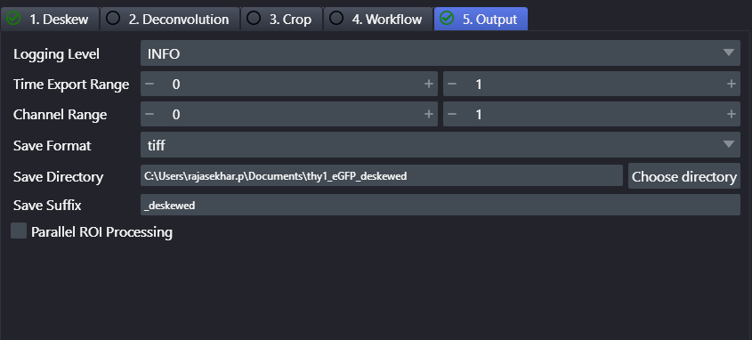
Option B — on the command line
Put the parameters in a YAML config and call lls-pipeline. This is the config shipped
in each dataset folder, with paths generalised to placeholders.
brain_organoid_config.yml
input_image: "/path/to/brain_organoid/EXP_7_5mm_2um_Steps_1_MMStack_Pos0_14.ome.tif"
save_dir: "/path/to/output/brain_organoid"
save_type: "tiff"
save_name: "brain_organoid_deskewed"
skew: "Y"
angle: 45
coverslip_rotation: False       # shear-only OPM geometry
invert_scan_direction: True     # scan runs opposite to the Zeiss LLS
physical_pixel_sizes:
  Z: 2.0
  Y: 1.04
  X: 1.04
thy1_eGFP.yml
input_image: "/path/to/thy1_eGFP/thy1gfp_3_8bit_250um_50fps_20ms_5s_10_MMStack_Pos0.ome.tif"
save_dir: "/path/to/output/thy1_eGFP"
save_type: "tiff"
save_name: "thy1_eGFP_deskewed"
skew: "Y"
angle: 45
coverslip_rotation: False       # shear-only OPM geometry
invert_scan_direction: True     # scan runs opposite to the Zeiss LLS
physical_pixel_sizes:
  Z: 1
  Y: 1
  X: 1
Run either config with:
Matching a different OPM system
Most OPM/SOPi acquisitions can be matched by combining three options — --skew X/--skew Y,
--no-coverslip-rotation, and --invert-scan-direction. If your first deskew looks
wrong, work through them in that order: skew axis, then coverslip rotation, then scan
direction.
Data availability and citations
The two example datasets are redistributed with the napari-lattice manuscript on Zenodo
(Supplementary_opm_data.zip), each with its raw image, deskewed output, and
lls-pipeline config. They originate from the following publications — please cite the
original sources if you use them:
-
brain_organoid— 4×-expanded brain organoid, direct-view oblique plane microscopy. Lamb, J. R., Cardoso Mestre, M., Lancaster, M., & Manton, J. D. (2025). Direct-view oblique plane microscopy. Optica, 12(4), 469–472. https://doi.org/10.1364/OPTICA.558420. Raw data: figshare 10.6084/m9.figshare.28324301. -
thy1_eGFP— uncleared coronal Thy1-GFP mouse brain section, scanned oblique plane illumination (SOPi). Kumar, M., & Kozorovitskiy, Y. (2019). Tilt-invariant scanned oblique plane illumination microscopy for large-scale volumetric imaging. Optics Letters, 44(7), 1706–1709. https://doi.org/10.1364/OL.44.001706. Raw data: zenodo.org/records/5088089.